
Coronary Angiography Indications.

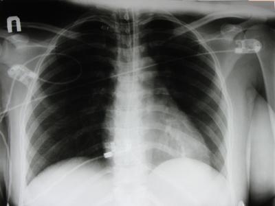
A coronary angiography, or coronary angiogram, is an X-ray examination of the arteries that supply the heart with blood. Usually these arteries are not visible on an X-ray, but for a coronary angiography a special dye is injected into the vessels to make them appear on an X-ray.This test can involve complications and should only be administered to patients who require it. Knowing the proper indications for when this procedure should be performed helps doctors determine which patients should undergo a coronary angiography.
Chest Pain
Chest pain, or angina, is an indication for coronary angiography. Chest pain can be a symptom of coronary artery disease. The angiogram allows doctors to visualize the condition of the coronary artery. They can look for any damage, narrowing, or obstruction that may be present within the blood vessel. The chest pain is usually located below the sternum and can either be a sharp or crushing pain. Patients also notice that pain worsens upon exertion or strenuous physical activity. The diseased coronary arteries inadequately supplying the heart with blood can be a cause of the chest pain.
Heart Failure
Patients who are experiencing heart failure due to unexplained reasons may need to undergo a coronary angiography. Heart failure is characterized by the heart's inability to pump enough blood to the rest of the body. This can be caused by damage to the heart muscle, abnormalities of the heart valves or an infection of the heart. In some cases, heart failure can be due to coronary artery disease. Once the other causes of heart failure have been ruled out, a coronary angiography can be performed to establish coronary artery disease as the underlying cause behind the heart failure.
Pain in the Jaw, Neck or Arm
Unexplained pain the jaw, neck or arm can be an indication for a coronary angiography. The pain can be a referred pain from the heart and coronary arteries. The pain actually originates from the heart itself, but is felt in the patient's jaw, neck or arm. The left jaw, neck and arm are usually the most common sites of pain referred from the heart. This pain can be a symptom of diseases within the coronary arteries.
Aortic Stenosis
Aortic stenosis is the abnormal narrowing of a special valve in the heart. Blood from the heart is pumped into the aorta, the main artery leaving the heart and providing blood to the rest body. In order to reach the aorta, the blood must pass through the aortic valve. If this valve does not open fully or becomes too narrow, then blood has a difficult time reaching the aorta. This narrowing of the valve is called aortic valve stenosis, or just aortic stenosis. Patients who are suspected of having aortic disease should undergo a coronary angiogram. A coronary angiogram can help determine the severity of the stenosis and can guide physicians in the selection of the proper treatment option.
What is a Dosimeter ?
Dosimeter is a device used to measure quantities of ionizing radiation, which includes both X-rays and radioactivity. Some types of dosimeters are used to measure exposure to other environmental factors as well, such as sound and ultraviolet rays. These devices come in a variety of shapes and sizes, and they are used primarily to monitor the amount of exposure to radiation in order to keep a person within safe levels.
Facts
The most common film dosimeters use what's called GafChromic film, which is a colorless film with a consistency of tissue paper. It develops a blue coloration if it encounters radiations. Film dosimeters are not reusable, but direct-reading devices, such as pocket dosimeters, can be reused. Thermoluminescents, or TLDs, are commonly used for medical purposes and gets its name because it thermally activates phosphorescence. TLDs are available in several different forms, including powders, chips, ribbons and rods.
History
The earliest known pocket dosimeter dates back to around 1932, and it was hand built by Charlie Lauritsen. One of the earliest film dosimeters was created by Ernest Wollan in 1943 to be used at the University of Chicago Metallurgical Laboratory. Thermoluminescent dosimeters were not used extensively until the 1960s when TLD badges became more popular. TLD ring badges were also developed in the late 1960s, and these small dosimeters were worn on a finger just like a regular ring.
Types
There are several types of dosimeters, including film, thermoluminescent, pocket, pocket chamber and others. Film and thermoluminescent dosimeters are usually worn as badges, rings or wrist bands. Pocket dosimeters are characterized as direct-reading devices, and pocket chambers are referred to as indirect-reading pocket dosimeters; the two are very similar in design and functionality. Pocket dosimeters are approximately the size and shape of a pen, and as its name implies, they fit easily inside of a pocket.
Function
Each type of dosimeter may function slightly differently, but they all serve the same purpose of detecting radiation or other environmental factors. A typical dosimeter body badge contains a substance, such as aluminum oxide, that is sealed inside a water-resistant packet. When exposed to radiation, the trapped electrons inside of the packet get excited and visible light is generated. The amount of light can then be measured to determine if the amount of radiation exposure is safe or not.
Size
Dosimeters are available in various shapes, sizes and forms. Some dosimeters, such as pocket models, are about the size of a pen and fit directly into your pocket. Other dosimeters devices are the size of badge and can be clipped onto a shirt. Film dosimeters come in a wide range of sizes from the size of a stamp to the size of a poster or even larger. Some thermoluminescent dosimeters are simply in powder form rather than being a standalone device.
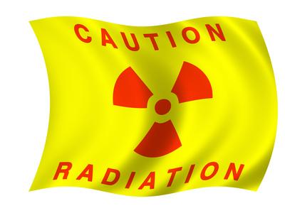
What are the effects of X-Ray Radiation on people ?

Approximately 15 percent of all natural and man-made types of radiation experienced from medical procedures. X-rays from these devices are measured in two ways, as entrance dose and effective dose. Entrance dose refers to the amount of radiation that enters the body, measured in units called grays, or Gy, and the effective dose is the response of absorption by specific tissues and organs.
Imaging
X-rays are used in diagnostic imaging to examine bones, teeth and internal structures. In CT scans, X-rays reveal cross-sections of the body, and in interventional radiology, X-rays direct internal procedures. X-rays pass through the body and are absorbed in differing amounts according to the density of the tissues and organs. The differing levels of exposure are captured on a detector, such as an X-ray film positioned behind the body part being radiated. This creates a picture that can then be examined.
Skin Injury
Skin damage can occur if patients are exposed to too much radiation from X-rays. The accepted amount of radiation before a patient may exhibit overexposure is 2Gy. "Radiology" states that the amount of exposure necessary to cause symptoms may vary from person to person based on numerous factors such as radiation sensitivity, the presence of diabetes mellitus or connective tissue disorders.
Fetal Birth Defects
The health of the mother weighed against the risk to the fetus must be taken into consideration in regards to X-ray imaging. It maintains that most radiation is not harmful to the fetus, especially if X-rays are of the extremities of the mother. In normal exposure of under 50 to 100 mGy, effects are none or so low as to be imperceptible. Over that level, spontaneous abortion, malformation, mental retardation or deficits in IQ may occur.
X-ray Radiation effects
X-rays are a kind of radiation, or electromagnetic wave. They’re very closely related to phenomena such as radio waves and waves of visible light, but unlike either of these, X-rays are capable of breaking bonds in biological molecules. This ability to destroy molecules denotes X-rays as a type of ionizing radiation, a category to which gamma radiation also belongs. Like gamma radiation, X-ray radiation effects can cause damage to living cells, tissues and organs. Damage can result from single high doses or can build up over time if many smaller exposures occur within a short period.
DNA Damage
DNA is genetic material. It’s a very long, delicate molecule that’s normally kept coiled up and protected in the nucleus of a cell. Dividing cells, however, uncoils DNA so that it can be replicated, and a copy passed to the new cell. That dividing cells are at much greater risk for damage from ionizing radiation such as X-rays than are cells that have their DNA protected. As such, rapidly-dividing cells, including skin cells, hair follicles and the lining of the digestive tract, are at greatest risk for damage.
Aging
Genetic material is not the only type of molecule at risk for breakage by X-rays. Proteins and structural tissues can become damaged as well. In particular, the collagen proteins that help skin maintain its elasticity are susceptible to damage; when collagen molecules break, they lose their ability to act as elastic skin supports. The U.S. Nuclear Regulatory Committee publishes a manual called “Biological Effects of Radiation” in which they note that aging effects of skin, organs and other tissues can result from X-ray exposure.
Cancer
Radiation exposure, even before birth, leads to increased risk for cancer. When genetic material within a cell becomes damaged, the damage often causes the cell to lose restrictions on its ability to grow and divide. As a result, damaged cells divide and grow rapidly, forming tumors that can steal nutrition from healthy body cells. This damages the organs in which the tumors are located, and can lead to organ failure and death. All cells are susceptible to genetic damage from X-rays, but the ones most likely to be affected are cells that are actively dividing when radiation exposure occurs.
Death
Exposure to large doses of radiation can overwhelm the body’s ability to heal damage. As such, very large single doses of X-rays or X-ray accumulation over time can lead to radiation sickness, including symptoms such as vomiting, disorientation and mental deficiencies. Depending upon the strength of the dose, radiation sickness can kill within minutes or up to several months after exposure.
