
Introduction to the Picture Archiving and Communication System
|

|
|
|
|
2nd Eb result sheet
| 02-171-2018results1911987913_1_.pdf |
|
Do you want to know ?
| ||
|
|

|
About Radiography
Radiography is the use of X-rays to view areas or objects that are normally not visible. X-rays are a form of radiation, focused into beams, which are able to pass through some materials and most types of tissue in the human body. Dense materials and tissue, such as bones, absorb X-rays and cause them to bounce back and appear white on film. This enables scientists, physicists, researchers, and other professionals to see inside the human body or detect hidden flaws in materials that would otherwise not be visible. The invention and advancement of radiography has greatly improved the diagnosis and treatment of medical conditions and has led to safer and more reliable manufacturing techniques.
Radiography Treatment
Radiography treatment is used to treat cancer. Radiography treatment is more commonly known as radiation therapy . It is the use of radiation as medical treatment, usually for forms of cancer. The therapeutic goal of radiation therapy, also known as radiotherapy, X-ray therapy and irradiation, is to kill cancer cells while doing as little damage as possible to normal cells. Unfortunately, it is never possible to completely avoid damaging normal cells during radiation therapy, so there will also be some degree of toxic effects from treatment.
Wilhelm Conrad Röntgen
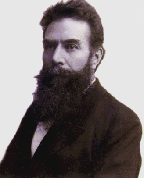
The 8th of November will be celebrated by radiographers worldwide as World Radiography Day to commemorate Wilhelm Conrad Roentgen’s discovery. On this day in 1895, Professor Roentgen was working with cathode rays using evacuated glass bulbs. He noted that when a current passed across the bulb, a barium platino-cyanide screen fluoresced and furthermore he noted the effect of the phenomenon on photographic plates. He termed this new discovery ‘X-Rays’. He further asserted that, with the use of these ‘X-Rays’ he was able to see through the body.Within three months of Roentgen’s discovery, radiographs were generated in major cities.
srt2007feb would like to encourage radiographers all over the world to celebrate this great discovery. World Radiography Days an annual international initiative intended to raise awareness of, and to stimulate an interest in, the profession of radiography. In addition it is aimed at highlighting the importance of radiography and the indispensable function it plays in the health care environment.
srt2007feb would like to encourage radiographers all over the world to celebrate this great discovery. World Radiography Days an annual international initiative intended to raise awareness of, and to stimulate an interest in, the profession of radiography. In addition it is aimed at highlighting the importance of radiography and the indispensable function it plays in the health care environment.
Mrs. Röntgen's hand, the first X-ray picture of the human body ever taken.
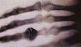
On 8 Nov, 1895, Wilhelm Conrad Röntgen (accidentally) discovered an image cast from his cathode ray generator, projected far beyond the possible range of the cathode rays (now known as an electron beam). Further investigation showed that the rays were generated at the point of contact of the cathode ray beam on the interior of the vacuum tube, that they were not deflected by magnetic fields, and they penetrated many kinds of matter. A week after his discovery, Rontgen took an X-ray photograph of his wife's hand which clearly revealed her wedding ring and her bones. The photograph electrified the general public and aroused great scientific interest in the new form of radiation. Röntgen named the new form of radiation X-radiation (X standing for "Unknown"). Hence the term X-rays (also referred as Röntgen rays, though this term is unusual outside of Germany).
Key Dates in Radiolgraphy History
1895 - Rontgen discovers x-rays.
1896 - Becquerel discovers radioactivity.
1901 - Rontgen receives the Nobel Prize in Physics for the discovery of x-rays.
1905 - The first English book on Chest Radiography is published.
1913 -Coolidge introduces the hot cathode tube.
1914 - Von Laue receives the Nobel Prize in Physics for x-ray diffraction from crystals.
1915 - Bragg and Bragg receive the Nobel Prize in Physics for crystal structure derived from x-ray diffraction.
1917 - Barkla receives the Nobel Prize in Physics for characteristic radiation of elements.
1918 - Eastman introduces radiographic film.
1920 - The Society of Radiographers is formed.
1924 - Siegbahn receives the Nobel Prize in Physics for x-ray spectroscopy.
1927 - Compton receives the Nobel Prize in Physics for scattering of x-rays by electrons.
1936 - Debye receives the Nobel Prize in Chemistry for diffraction of x-rays and electrons in gases.
1934 - Joliot and Curie discover artificial radionuclides.
1937 -The first clinical use of artificial radioactivity is done at the University of California- Berkeley.
1946 - Schoenander develops the film cassette changer which allowed a series of cassettes to be exposed at the rate of 1.5 cassettes per second.
1946 - Nuclear medicine is discovered by accident.
1950 's - Wide-spread clinical use of nuclear medicine starts.
1950's - Development of the image intensifier and X-ray television.
1956 - The medical use of Ultrasound starts in Poland.
1962 -Kuhl introduces emission reconstruction tomographyThis method later becomes known as SPECT and PET.
1967 - The first clinical use of MRI takes place in England.
1972 - CT is invented by British engineer Godfrey Hounsfield of EMI Laboratories in England.
1977 - The first human MRI images are produced.
1979 - Comack and Hounsfield receive the Nobel Prize in Medicine for computed axial tomography.
1980's - The advancement of radiopharmaceuticals and the use of computers transform Nuclear Medicine into what it is today.
1980's - Fuji develops CR technology.
1981 - Siegbahn receives the Nobel Prize in Physics for high resolution electron spectroscopy.
1984 - MRI is cleared for commercial use by the Food and Drug Administration.
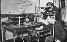
Taking an X-ray image with early Crookes tube apparatus, late 1800s.
|
| ||
Click to set custom HTML
