
Alternatives for Angiography
Angiography describes a diagnostic procedure that allows doctors to visualize the inside of blood vessels. Angiography can determine the blood flow in and around organs such as the lungs, brain and kidneys, but doctors commonly perform coronary angiography to examine blood flow through the heart and the heart's pumping ability. Although coronary angiography can eliminate the need for major heart surgery, as noted by the American College of Radiology, the risks lead some patients to search for alternative procedures.
Risks of Traditional Angiography To perform a traditional coronary angiography, doctors insert a long, thin, flexible tube known as a catheter through a small incision into an artery, usually in the arm or leg. The doctor then threads the catheter through the blood vessels until they find the coronary artery. They then inject a contrast medium, or dye, into the arteries to allow visualization through X-rays.Risks of traditional angiography include bleeding and infection at the site of the incision. Because a catheter enters the blood vessels, the risk of introducing infection into the bloodstream also increases. As the catheter travels through the blood vessels, it can puncture the vessel, causing internal bleeding. The catheter can also promote the formation of a blood clot that can result in a heart attack or stroke.
Magnetic Resonance Angiography
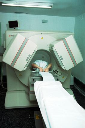
Magnetic resonance angiography uses magnetic resonance imaging--MRI--technology to visualize inside blood vessels. Unlike traditional angiography, doctors consider magnetic resonance angiography a noninvasive procedure.
Patients only need to lie on a table and slide into the tunnel-like machine for approximately one hour. The MRI machine uses magnets and radio waves to produce images of the inside of the body. To visualize blood vessels better, a contrast medium may be injected through a needle. This type of angiography is considered safe with few associated risks.
Multidetector Computed Tomography Angiography
Another non-invasive alternative to traditional angiography is the multi-detector computed tomography angiography, also called the multislice CT. This procedure uses high-energy radiation, similar to an X-ray, and 16 to 256 rows of detectors to visualize the images.
Like traditional angiography, multislice CT requires the use of a contrast medium to help make the blood vessels visible. Although the multislice CT utilizes higher doses of radiation than the X-rays of a traditional angiography, it presents fewer health risks to the patient. Multi-detector computed tomography provides the advantage of higher spatial resolution of the images with shorter and less invasive examinations.
Risks of Traditional Angiography To perform a traditional coronary angiography, doctors insert a long, thin, flexible tube known as a catheter through a small incision into an artery, usually in the arm or leg. The doctor then threads the catheter through the blood vessels until they find the coronary artery. They then inject a contrast medium, or dye, into the arteries to allow visualization through X-rays.Risks of traditional angiography include bleeding and infection at the site of the incision. Because a catheter enters the blood vessels, the risk of introducing infection into the bloodstream also increases. As the catheter travels through the blood vessels, it can puncture the vessel, causing internal bleeding. The catheter can also promote the formation of a blood clot that can result in a heart attack or stroke.
Magnetic Resonance Angiography
Magnetic resonance angiography uses magnetic resonance imaging--MRI--technology to visualize inside blood vessels. Unlike traditional angiography, doctors consider magnetic resonance angiography a noninvasive procedure.
Patients only need to lie on a table and slide into the tunnel-like machine for approximately one hour. The MRI machine uses magnets and radio waves to produce images of the inside of the body. To visualize blood vessels better, a contrast medium may be injected through a needle. This type of angiography is considered safe with few associated risks.
Multidetector Computed Tomography Angiography
Another non-invasive alternative to traditional angiography is the multi-detector computed tomography angiography, also called the multislice CT. This procedure uses high-energy radiation, similar to an X-ray, and 16 to 256 rows of detectors to visualize the images.
Like traditional angiography, multislice CT requires the use of a contrast medium to help make the blood vessels visible. Although the multislice CT utilizes higher doses of radiation than the X-rays of a traditional angiography, it presents fewer health risks to the patient. Multi-detector computed tomography provides the advantage of higher spatial resolution of the images with shorter and less invasive examinations.
Advantages & Disadvantages of a CT Scan
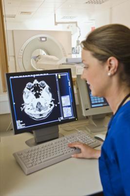
CT scanning is a highly accurate medical test that "combines special x-ray equipment with sophisticated computers to produce multiple images or pictures of the inside of the body. While this painless and noninvasive procedure greatly assists radiologists in diagnosing cardiovascular diseases, musculoskeletal problems, infectious diseases, trauma and certain types of cancer, the CT scan also carries radiation-induced risks. The San Francisco Examiner reports that over 60 million CT scans are performed each year in the United States. Here are some pros and cons of this sophisticated technology.
Comprehensiveness The CT scan can record images of bone, soft tissue and blood vessels simultaneously, offering clear advantages over standard x-rays. The CT scans' diagnostic ability can make surgical biopsy or exploratory surgery unnecessary. Its real-time imaging allows CT scanning to be used to guide needle biopsies and similar procedures. A cardiologist can view "clear 3D images of the coronary arteries without having to do an invasive angiography.
Speed
Most partial CT scans take just seconds, and a full scan of the entire body can be completed in about a half-hour. This speed helps in finding internal injuries and internal bleeding quickly enough to help save lives.
Fewer Restrictions than MRI
CT scans have a less stringent requirement for patients to remain absolutely motionless during the scan as compared to Magnetic Resonance Imaging (MRI). CT scans, unlike MRI, have no prohibition on implanted medical devices.
Concerns over Radiation
An international study found that levels of radiation emitted by CT scanners vary widely. At maximum, "patients can be getting radiation equal to 600 chest x-rays," leading to the possibility of lung cancer or breast cancer as a consequence. X-rays also damage DNA itself. Since the federal government has no regulations limiting how much radiation patients may receive during CT scans, the referring physician decides the strength of the dose. Some authorities estimate that CT scans cause one percent of cancers in America. According to the Examiner, patients who have had an abdominal CT scan run a cancer risk of one in 1,000.
Unsuitability for Pregnant Women
CT scans are, "in general, not recommended for pregnant women unless medically necessary because of potential risk to the baby." Women should let their doctor and scan technician know if there is any chance they are pregnant. Additionally, whether pregnant or not, women face a higher risk than men since radiation can damage their ovaries. The San Francisco Examiner reports that women in their twenties run a breast cancer risk of approximately one percent from chest CT scans.
Dangers to Children
Nursing babies are potentially at risk from "contrast material" injected into their mothers before CT scans and a 24-hour waiting period is recommended before resuming breast-feeding. Children are also highly sensitive to radiation and so CT scans should not be done on children without absolute need.
Overuse of CAT Scans
As many as one-third of CT scans "might not be justified," since overuse of this type of testing increases cancer risks for patients. The New England Journal of Medicine linked this overuse of CAT scans to nearly 2 percent of all projected U.S. cancer diagnoses over the next 20 to 30 years.
Next Generation PDT / Cancer Treatment For The 21 st Century
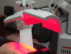
Next Generation PDT (NGPDT) has developed a uniquely effective Photodynamic Therapy (PDT) for the treatment of most cancers. By building on proven and existing medical research for PDT cancer treatments, Next Generation PDT is successfully treating a wide variety of cancers non-invasively and with greater effect than conventional therapies.
NGPDT has been achieving excellent results through the development of a new generation photosensitizer which can identify and selectively accumulate within cancerous tumour tissues. Our advanced light and laser delivery methods can activate the abosrbed photosensitiser within both surface and deep seated tumours. Next Generation PDT is a safe, pain free cancer therapy, without the prolonged photosensitivity concerns introduced by traditional PDT protocols.
In combination with our advanced 'next generation' photosensitizer, NGPDT's whole-body light delivery methods allow for the effective treatment of both deep seated and metastatic cancers. PDT provides an effective means of treatment without the potentially debilitating and health ravaging therapys, or invasive surgery usually associated with cancer. TREATING A WIDE RANGE OF CONDITIONS We have had great success in treating a wide range of cancer conditions over the last 7 years. Each of our patients is individually assessed by qualified specialists prior to prescribing any kind of treatment protocol.
Prostate Cancer Breast Cancer Bladder Cancers
Colorectal Cancers Brain Tumours Soft Tissue Sarcomas
Germ Cell Tumours Retinoblastoma Kidney Cancers
Lung Cancer Liver Cancer And Others
