
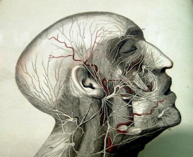
Diagnostic Radiography
Diagnostic Radiography refers to the controlled use of ionising radiation or radioactive substances and non-ionising radiation and substances to facilitate the diagnosis, management and treatment of disease or injury.† Diagnostic Radiography modalities that use ionizing radiation or radioactive substances include Projection Radiography, Fluoroscopy, Image Intensifier (II), Computed Tomography (CT), Angiography or Vascular Interventional Radiology (VIR), Nuclear Medicine (NM), Positron Emission Tomography (PET), Bone Mineral Densitometry (BMD), and other technologies that may be introduced. Modalities that use non-ionizing radiation and substances include Ultrasound (US), Magnetic Resonance Imaging (MRI), Extracorporeal Shockwave Lithotripsy (ESWL), and other technologies that may be introduced.
Below is a simple and comprehensive video that explains how does an X-ray Tube work.
Radiography technique
* CXR - Chest X-Ray
* IVU (Intravenous Urogram)
* ERCP (Endoscopic Retrograde Cholangio pancreatography)
* Micturating Cystourethrogram (MCUG)
* CXR - Chest X-Ray
* IVU (Intravenous Urogram)
* ERCP (Endoscopic Retrograde Cholangio pancreatography)
* Micturating Cystourethrogram (MCUG)
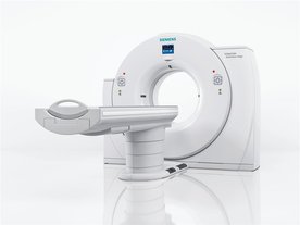
Computed Tomography (CAT scans, CT scans)
Computed tomography, also know as a CT, is a specialized form of X-ray. In this test, you will lay on a table that faces a special array that looks like a large donut. When the test starts, the table will move through the donut as the images are collected. This test creates cross-sectional images that a radiologist is able to scroll through to get detailed images of internal organs. CT scans are exceptional tools for emergency procedures because they are quick. A full-body CT scan can be completed in a matter of minutes, and it is effective for looking at organs and discerning whether there is internal bleeding. In certain instances, contrast material can be given by mouth or in a vein through an IV so that certain vascular or gastrointestinal structures can be better visualized.
Computed tomography, also know as a CT, is a specialized form of X-ray. In this test, you will lay on a table that faces a special array that looks like a large donut. When the test starts, the table will move through the donut as the images are collected. This test creates cross-sectional images that a radiologist is able to scroll through to get detailed images of internal organs. CT scans are exceptional tools for emergency procedures because they are quick. A full-body CT scan can be completed in a matter of minutes, and it is effective for looking at organs and discerning whether there is internal bleeding. In certain instances, contrast material can be given by mouth or in a vein through an IV so that certain vascular or gastrointestinal structures can be better visualized.
Magnetic Resonance Imaging (MRI)
Magnetic resonance imaging (MRI) is similar in nature to a CT scan in that the images obtained are cross-sections of the body. When having an MRI, you must lay flat on a table that will move you into and out of a large tube. This tube is constricting, and patients that suffer from anxiety have difficulty with MRIs, which take an hour or more to complete. But there are some distinct advantages to putting up with this test, as the image quality is excellent. MRIs are exceptional for looking at soft tissue structures, such as the brain. An MRI can often tell days before a CT whether a patient has suffered a stroke, because of its ability to distinguish between different densities. Back to top
Magnetic resonance imaging (MRI) is similar in nature to a CT scan in that the images obtained are cross-sections of the body. When having an MRI, you must lay flat on a table that will move you into and out of a large tube. This tube is constricting, and patients that suffer from anxiety have difficulty with MRIs, which take an hour or more to complete. But there are some distinct advantages to putting up with this test, as the image quality is excellent. MRIs are exceptional for looking at soft tissue structures, such as the brain. An MRI can often tell days before a CT whether a patient has suffered a stroke, because of its ability to distinguish between different densities. Back to top
Cerebral Angiography Procedures
Doctors perform a diagnostic procedure known as an angiography, also called an arteriography, to visualize the blood vessels in a particular area of the body. A cerebral angiography provides images of the blood vessels in and around the brain. This test can reveal abnormalities in the blood vessels such as a bulge, or aneurysm, inflammation, or vasculitis, or the presence of a blood clot. Although the risk of complications remains small, following a specific set of procedures helps to alleviate patient anxiety before, during and after the test.
Prior to Procedure
As with any medical procedure, the patient must sign a consent form. Patients should also alert their doctors if they have any allergies to shellfish or bleeding problems, have experienced an allergic reaction to contract dye in the past or are currently pregnant. Doctors may perform routine tests on blood and urine samples prior to the procedure.
The doctor will instruct the patient to refrain from eating or drinking for a period of time prior to the start of the test. Once at the testing site, the patient will remove all clothing and jewelry and put on a hospital gown. Most patients will receive an IV so doctors can administer fluids and medications for pain and anxiety.
Catheter Insertion
To begin, doctors insert a long, thin, flexible tube known as a catheter through a small incision in an artery. Doctors often insert through the artery in the groin, but may also use an artery in the arm, neck or thigh, according to Atlanta Brain and Spine Care. A local anesthetic helps to numb the area to reduce pain.
The doctor then carefully moves the catheter through the main blood vessels to the abdominal and chest region and into the artery of the neck. X-ray images taken during the procedure help guide the catheter.
Injection of Dye
Once the catheter has reached the carotid artery, doctors inject a contrast dye through the catheter. The contrast dye usually consists of iodine and water and helps change the opacity of the blood vessels so they appear clearer on the X-ray images. Doctors can then see how the contrast dye moves through the blood vessels, which can reveal the presence of any blockages. Patients may feel a warming or burning sensation as doctors inject the dye.
Imaging
After injecting the dye, doctors use high energy radiation--X-rays--to produce images. As the X-rays pass through the body they are absorbed at different levels, allowing the image to differentiate between the different tissues. A camera detects these images and displays them as movie of the blood flow.
Following the Procedure
At the completion of the test doctors, remove the catheter, bandage the incision and observe the patient while they remain lying still. Once doctors allow the patient to get up and move around the patient may feel lightheaded or dizzy, especially if they get up too quickly.
Back to top
Types of iodine Contrst Used in CT Scan
CT scans are a radiographic test used to identify heart disease, blood clots, broken bones and cancers quickly and painlessly. Also known as computed tomography, the CT scanner takes pictures in slices, providing a transaxial view of all soft tissues and organs. An iodine contrast agent, or contrast media, is frequently used to increase visualization of the desired tissues. Although many brands of iodinated contrast are available, three main types of iodine contrast are administered routinely.
Intravenous
Intravenous iodine contrast is employed when the ordering physician needs to see specific blood vessels or highly vascular organs. The liver, brain, spinal cord, heart and kidneys might require use of intravenous contrast for better visualization. Some people may experience a metallic taste in their mouths immediately following an iodine contrast injection.
Oral
Several iodine suspensions are suggested for enteral use to view the esophagus, stomach or small intestine. Commonly used in the United States, gastrografin is chemically altered for palatability. Gastrografin is a yellowish liquid that contains a lemon-like flavor. Some people complain of difficulty ingesting gastrografin due to its bitter taste. Gastrografin can be used in conjunction with intravenous iodine to highlight all abdominal structures.
Rectal
Orally ingested iodine will eventually travel to the colon; however, some of the iodine element may break down, resulting in a poor picture. For radiographic CT images of the colon and lower intestine, an iodine agent must be administered rectally via an enema. Using a regular enema bag, a nurse or technician inserts the ordered amount of iodine contrast into the colon through the anus. A feeling of fullness or diarrhea can result from rectally administered iodine contrast. Back to top
